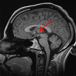
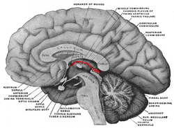
Human brain left midsagitttal view closeup description 2
John A Beal, PhD
Dep't. of Cellular Biology & Anatomy, Louisiana State University Health Sciences Center ShreveportHuman brain left - midsagitttal view - closeup
- Corpus callosum, Rostrum
- Corpus callosum, Genu
- Corpus callosum, Corpus
- Corpus callosum, Splenium
- Septum pellucidum
- Fornix, Corpus
- Glandula epiphysialis
- Recessus pinealis
- Habenula
- Stria medullaris thalami
- Thalamus (Pars dorsalis)
- Adhaesio interthalamica
- Plexus choroideus
- Foramen interventriculare
- Comissura anterior
- Hypothalamus
- Lamina terminalis
- Recessus supraopticus
- Recessus infundibuli
- Infundibulum
- Tuber cinerum
- Corpora mamillaria
- Sulcus hypothalamicus (blue line)
- Mesencephalon (Crus cerebri)
- Tegmentum mesencephali
- Aqaeductus mesencephali
- Tectum mesencephali, a) Colliculus superior B) C. inferior
- Pons
- Ventriculus quartus
- Cerebellum
- Comissura posterior
The Thalamus (Diencephalon) is part of the Forebrain and is divided into Dorsal Thalamus (Superior), Hypothalamus (Inferior and Medial) and Subthalamus (Lateral to Hypothalamus).
On a half brain specimen, the Thalamus can be identified. The Thalamus (anteroom) is connected caudally with the midbrain and rostrally with the cerebral hemispheres. Note that the walls of the 3rd ventricle are completely formed by the Thalamus. The Hypothalamic Sulcus separates the Dorsal Thalamus superiorly from the Hypothalamus inferiorly. The subthalamus is located lateral to the hypothalamus and is not visible here.
The anterior wall of the 3rd ventricle is a thin sheet of tissue called the Lamina Terminalis. In the Dorsal Thalamus note the Interthalamic Adhesion (Massa Intermedia), and the three parts of the Epithalamus: Stria Medullaris Thalami, Habenula, and the Pineal Gland. The floor of the Hypothalamus is made up of the Infundibulum, which connects with the pituitary gland, and posteriorly, the Tuber Cinereum and Mammillary Body.
Relevantní obrázky
Relevantní články
ThalamusThalamus je spolu s epithalamem součástí zadní části mezimozku (diencephalon) a je seskupením senzorických, asociačních a nespecifických jader. Zprostředkovává převod informací přicházejících z periférie do specifických projekčních a asociačních oblastí mozkové kůry a do důležitých center mozečku. Umožňuje také vzájemnou interakci vyšších oddílů CNS. .. pokračovat ve čtení
EpithalamusEpithalamus je jedním z oddílů mezimozku (diencephalon). .. pokračovat ve čtení


